LETTER TO THE EDITOR
Doi: 10.5578/tt.10463
Tuberk Toraks 2016;64(3):256-257

Let's get estimates!
Ayşe G?l ?NEY KURNAZ1, Dilek ŞEN DOKUMACI2, Funda YAL?IN1, Turgay ULAŞ3, Mehmet GENCER1
1 Department of Chest Diseases, Faculty of Medicine, Harran University, Sanliurfa, Turkey
1 Harran ?niversitesi Tıp Fak?ltesi, G?ğ?s Hastalıkları Anabilim Dalı, Şanlıurfa, T?rkiye
2 Division of Radiology, Faculty of Medicine, Harran University, Sanliurfa, Turkey
2 Harran ?niversitesi Tıp Fak?ltesi, Radyoloji Bilim Dalı, Şanlıurfa, T?rkiye
3 Division of Hematology, Faculty of Medicine, Harran University, Sanliurfa, Turkey
3 Harran ?niversitesi Tıp Fak?ltesi, Hematoloji Bilim Dalı, Şanlıurfa, T?rkiye
A 57-year-old female patient with hairy cell leukemia was consulted to us to evaluate her routine chest radiograph. The patient had no symptomand her chest physical examination findings were normal. PA chest X-Ray revealed linear, weak sided, crinkly opacity at right hemithorax and 4 x 2.5 cm, well circumscribed opacity at left retrocardiac region (Figure 1). It was thought that the lesion on the left chest might be a mass and the pathology on right chest could not be fully described. Is it pleural plaque due to asbestos? No, because the inner margin is often well defined while the tapering outer margin is indistinct in pleural? plaque but both of sides are indistinct on our patient's chest X-Ray. Is it cavity? No, because cavity means for surrounded by parenchima of the lung but the upper side of this lesion was not seen. Is it foreign body like a necklace? No, because we had already knew that there was no foreign body on the patient. Then what you should think about this lesion?

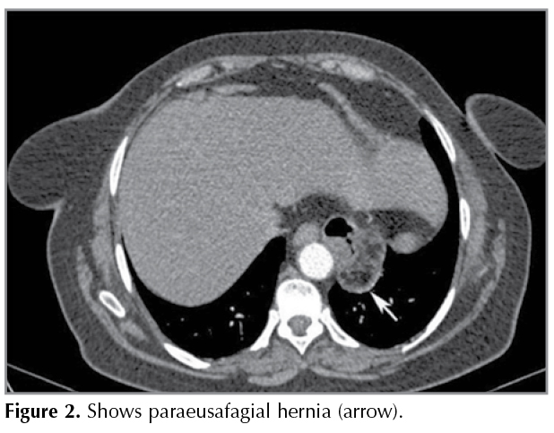
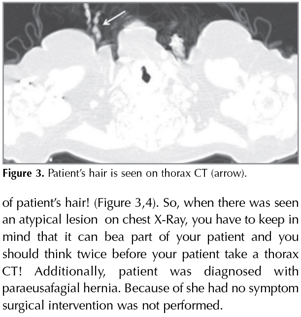
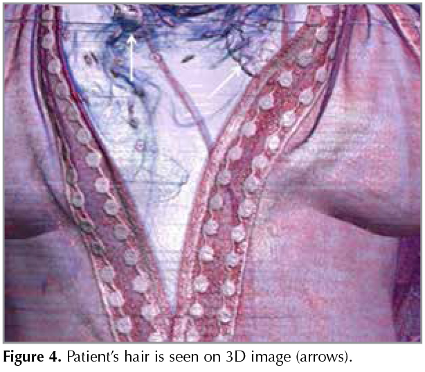
Thorax computed tomography (CT) was taken to better identify the lesion on right chest and retrocardiac opacity. Thorax CT showed paraeusafagial hernia at left side of diaphragm (Figure 2). The pathology seenon right hemithorax on chest X-Ray was described as plait of patient's hair! (Figure 3,4). So, when there was seen an atypical lesion? on chest X-Ray, you have to keep in mind that it can bea part of your patient and you should think twice before your patient take a thorax CT! Additionally, patient was diagnosed with paraeusafagial hernia. Because of she had no symptom surgical intervention was not performed.
Yazışma Adresi (Address for Correspondence)
Dr. Ayşe G?l ?NEY KURNAZ
Harran ?niversitesi Tıp Fak?ltesi,
G?ğ?s Hastalıkları Anabilim Dalı,
ŞANLIURFA - TURKEY
e-mail: ayseguloney81@hotmail.com
Geliş Tarihi/Received: 29.12.2015 - Kabul Ediliş Tarihi/Accepted: 07.01.2016