LETTER TO THE EDITOR
Doi: 10.5578/tt.9972
Tuberk Toraks 2015;63(3):210-211

Pseudotumor by lateral process of the vertebra
Tomohiro TAMURA1, Gen OHARA1, Katsunori KAGOHASHI1, Hiroaki SATOH1
1 Division of Respiratory Medicine, Mito Medical Center, Tsukuba University, Mito, Japan
1 Tsukuba ?niversitesi Mito Tıp Merkezi, Solunum Bilim Dalı, Mito, Japonya
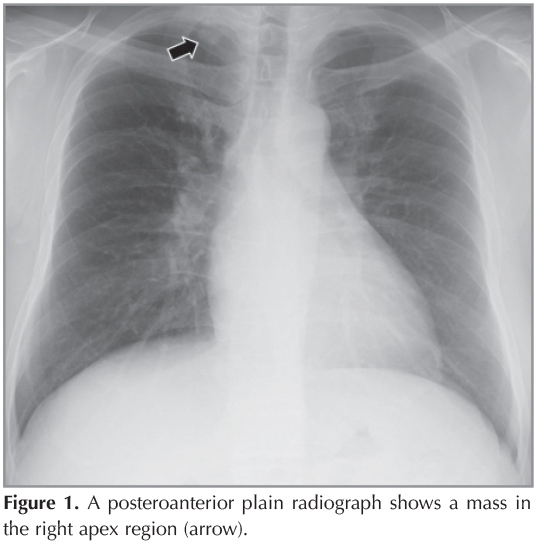
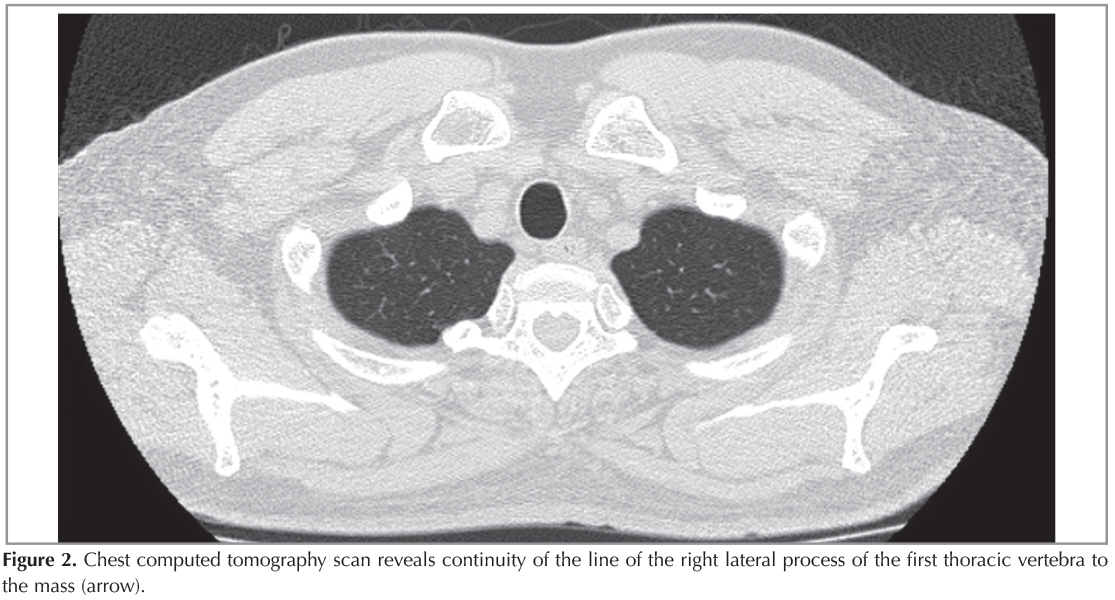
A 46-year-old man with no symptom was referred for a mass in the right apex incidentally noted on chest radiograph (Figure 1). He had no medical history. The axial view of chest computed tomography (CT) scan revealed continuity of the line of the right lateral process of the second thoracic vertebra to the mass no other abnormality in lung and thorax was found (Figure 2,3). Therefore, the patient was diagnosed to have anatomical variation of right lateral process of the thoracic vertebra. As the patient had no symptom, surgical resection was not performed.

Anatomic variations of thorax can simulate pathologic lesions on plain posteroanterior radiographs (1). Ossification of the first rib head is known to produce findings on chest radiographs that can be mistaken for lung tumor (1,2). Chest radiograph is a pivotal role as the first-line for detection of various kinds of chest diseases, although anatomical variations or superimposition of normal structures to form a composite opacity can mimic a lesion. Especially in pulmonary apex region, the images of CT scan is characteristic enough for making a definitive diagnosis and render other diagnostic modalities unnecessary. Although rare, physicians should be taken this anatomic variation into differential diagnosis in case with apex mass lesion.
CONFLICT of INTEREST
None declaared.
REFERENCES
- Gronner AT, Ominsky SH. Plain film radiography of the chest: findings that simulate pulmonary disease. AJR Am J Roentgenol 1994;163:1343-8.
- Fakhry SM, Thomas CG Jr. Pseudotumor of the supraclavicular fossa. South Med J 1986;79:822-4.
Yazışma Adresi (Address for Correspondence)
Dr. Hiroaki SATOH
University of Tsukuba, Division of Respiratory Medicine,
Mito Medical Center, MITO - JAPAN
e-mail: hirosato@md.tsukuba.ac.jp