SHORT REPORT
Doi: 10.5578/tt.8132
Tuberk Toraks 2015;63(2):109-110

?leri ya?ta bir olguda Scimitar sendromuna ait ?KBT bulgular?
Canan ALTAY1, Naciye Sinem GEZER1, I??l BA?ARA1, P?nar BALCI1, Ey?p Sabri U?AN2
1 Dokuz Eyl?l ?niversitesi T?p Fak?ltesi, Radyoloji Anabilim Dal?, ?zmir, T?rkiye
1 Department of Radiology, Faculty of Medicine, Dokuz Eylul University, Izmir, Turkey
2 Dokuz Eyl?l ?niversitesi T?p Fak?ltesi, G???s Hastal?klar? Anabilim Dal?, ?zmir, T?rkiye
2 Department of Chest Diseases, Faculty of Medicine, Dokuz Eylul University, Izmir, Turkey
?ZET
?leri ya?ta bir olguda Scimitar sendromuna ait ?KBT bulgular?
Scimitar sendromu sa? akci?er ven?z d?n???n?n anormal olarak inferior vena kavaya oldu?u nadir bir vask?ler anomalidir. Biz burada ileri ya?ta tan? alan bir Scimitar sendromlu olguyu sunmaktay?z.
Anahtar kelimeler: Scimitar sendromu, ?KBT
SUMMARY
MDCT findings of Scimitar syndrome in an elderly patient
Scimitar syndrome is a rare congenital vascular anomaly characterized by abnormal venous drainage of the right lung to the inferior vena. We report here a case who is diagnosed late with Scimitar syndrome.
Key words: Scimitar syndrome, MDCT
INTRODUCTION
A 68-year-old woman was referred to our radiology department from chest diseases clinic with suspicion of sarcoidosis. She complained subfebrile fever and cough of a few months duration without laboratory and radiologic findings of infection. On detailed questioning, there was no history of any hemoptysis, expectoration, chest pain and extrapulmonary symptoms.
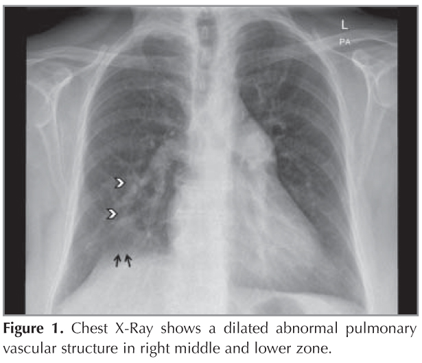
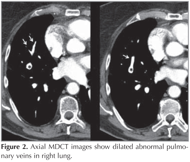
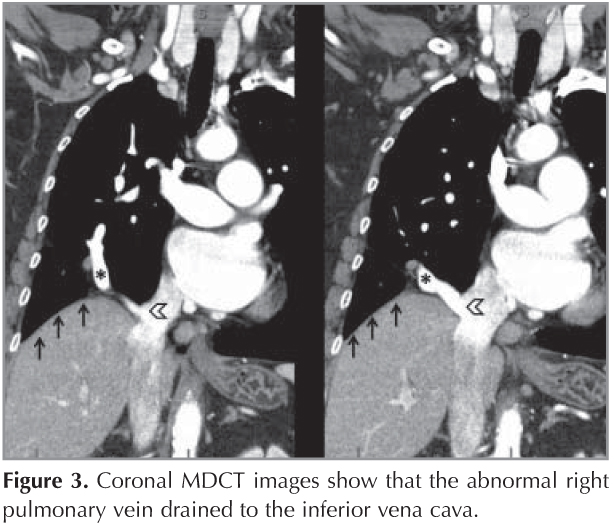
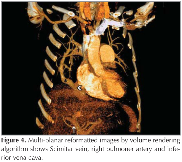
Chest X-ray showed a dilated abnormal pulmonary vascular structure (arrowheads) in right middle and lower zone (Figure 1). There was also seen irregularity of the medial right hemidiaphragm (short arrows). Axial image of contrast enhanced thorax multi detector computed tomography (MDCT) showed dilated right superior pulmonary vein (RSPV) (Figure 2, asterisk). The middle pulmonary vein also drained into RSPV (short arrows). Coronal reformatted MDCT images revealed mild right lung hypoplasia, elevation of right hemidiaphragm (short arrow) and draining the RSPV (asterisk) to the vena cava inferior (VCI) (arrowhead) (Figure 3). Volume rendering MDCT image showed relationship between RSPV (asterisk) with VCI (arrowhead) (Figure 4). There was also seen right pulmonary artery (dagger) and portal vein (short arrow). Final diagnosis of the patient was partial anomalous right pulmonary venous connection (Scimitar syndrome).
Scimitar syndrome is an uncommon vascular pathology of lung characterized by partial anomalous right pulmonary venous connection of the right pulmonary veins (1). The dilated right pulmonary vein called also "Scimitar" and usually drained to the infra-diaphragmatic VCI or hepatic veins. Scimitar syndrome has two varieties; the infantile form presents heart failure with other associated cardiac anomalies in first life of life. The adult form is usually asymptomatic and not associated with other cardiac disorders (2). The major clinic findings are hemoptysis and pulmonary arterial hypertension. Additionally, right lung hypoplasia, dextrocardia and anomalous systemic arterial supply from aorta to the right lung may be isolated.
CONFLICT of INTEREST
None declared.
REFERENCES
- Siu CW, Cheung SC, Chan CW, Lam YM, Tse HF, Jim MH. Scimitar syndrome on chest X ray. Postgrad Med J 2008;84:558. doi:10.1136/pgmj.2008.070375.
- Frydrychowicz A, Landgraf B, Wieben O, Fran?ois CJ. Images in Cardiovascular Medicine. Scimitar syndrome: added value by isotropic flow-sensitive four-dimensional magnetic resonance imaging with PC-VIPR (phase-contrast vastly undersampled isotropic projection reconstruction). Circulation 2010;121:e434-6.
Yaz??ma Adresi (Address for Correspondence)
Dr. Canan ALTAY
Dokuz Eyl?l ?niversitesi T?p Fak?ltesi,
Radyoloji Anabilim Dal?,
Mithatpa?a Cad.
?nciralt? ?ZM?R - TURKEY
e-mail: cananaltay@yahoo.com