ED?T?RE MEKTUP
Doi: 10.5578/tt.8064
Tuberk Toraks 2015;63(1):65-66

Orta ya?l? bir hastada Bochdalek hernisi
Hiroko WATANABE1, Tomohiro TAMURA1, Kesao IGUCHI2, Katsunori KAGOHASHI1, Hiroaki SATOH1
1 Tsukuba ?niversitesi Mito T?p Merkezi, Solunum Hastal?klar? Anabilim Dal?, Ibaraki, Japonya
1 Department of Respiratory Medicine, Mito Medical Center, Tsukuba University, Ibaraki, Japan
2 Tsukuba ?niversitesi Mito T?p Merkezi, G???s Cerrahisi Anabilim Dal?, Ibaraki, Japonya
2 Department of Thoracic Surgery, Mito Medical Center, Tsukuba University, Ibaraki, Japan
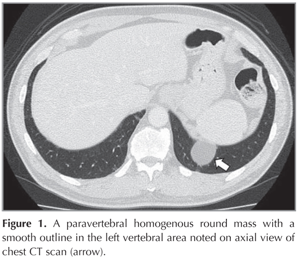
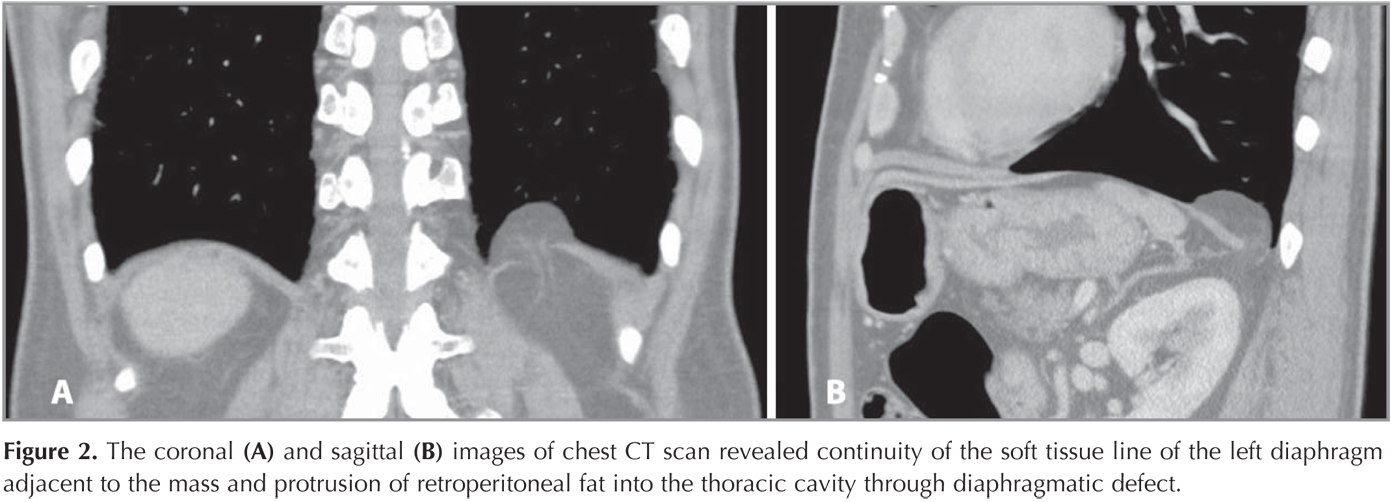
A 59-year-old man with no symptom was referred for a paravertebral homogenous round mass with a smooth outline in the left vertebral area incidentally noted on axial view of chest computed tomography (CT) scan (Figure 1). He had no medical history. The coronal and sagittal images of chest CT scan revealed continuity of the soft tissue line of the left diaphragm adjacent to the mass and protrusion of retroperitoneal fat into the thoracic cavity through diaphragmatic defect (Figure 2). Therefore, the patient was diagnosed to have Bochdalek hernia. As the patient had no symptom and benign nature of the disease, surgical resection was not performed.
Bochdalek hernia is one of the most common types of diaphragmatic hernia (1). Chest radiograph is a pivotal role as the first-line for detection of various kinds of chest diseases, although imaging for diagnosis of the disease is mainly based on conventional axial sections of CT scan. In these images, diaphragmatic hernias were sometimes misdiagnosed as other diseases in lung parenchyma (2,3). The coronal and sagittal images of CT scan as well as? magnetic resonance imaging (MRI) for a Bochdalek hernia are characteristic enough for making a definitive diagnosis and render other diagnostic modalities unnecessary (4,5). Although very rare, physicians should be taken this disease into differential diagnosis in case with paradiaphragmatic mass lesion.
CONFLICT of INTEREST
None declared.
Geli? Tarihi/Received: 22.07.2014 • Kabul Edili? Tarihi/Accepted: 08.08.2014
REFERENCES
- Schumacher L, Gilbert S. Congenital diaphragmatic hernia in the adult. Thorac Surg Clin 2009;19:469-72.
- Snyder HS, Salo DF, Kelly PH. Congenital diaphragmatic hernia presenting as massive gastrothorax. Ann Emerg Med 1990;19:562-4.
- Somani SK, Gupta P, Tandon S, Sonkar D, Bhatnagar S, Saxena M. Bochdalek diaphragmatic hernia masquerading as tension hydropneumothorax in an adult. J Thorac Cardiovasc Surg 2011;141:300-1.
- Shin MS, Mulligan SA, Baxley WA, Ho KJ. Bochdalek hernia of diaphragm in the adult. Diagnosis by computed tomography. Chest 1987;92:1098-101.
- Sugimura A, Kikuchi J, Satoh M, Ogata M, Inoue H, Takishima T. Bilateral Bochdalek hernias in an elderly patient diagnosed by magnetic resonance imaging. Intern Med 1992;31:281-3.
Yaz??ma Adresi (Address for Correspondence)
Dr. Hiroaki Satoh
Tsubaka ?niversitesi Mito T?p Merkezi,
Solunum Hastal?klar? Anabilim Dal?,
Miya-Machi 3-2-7, Mito 310-0015,
IBARAKI - JAPAN
e-mail: hirosato@md.tsukuba.ac.jp