LETTER TO THE EDITOR
Doi: 10.5578/tt.7866
Tuberk Toraks 2014;62(4):322-323

Kemik ili?i tutulumu ve s?k tekrarlayan bilateral pn?motoraks ile seyreden bir milier t?berk?loz olgusu
Abdullah ??M?EK1, M?jgan G?LER1, H?lya ?ELENK ERG?DEN1, Ruhsar OFLUO?LU1, Nermin ?APAN1
1 Ankara Atat?rk G???s Hastal?klar? ve G???s Cerrahisi E?itim ve Ara?t?rma Hastanesi, G???s Hastal?klar? Klini?i,
Ankara, T?rkiye
1 Department of Chest Diseases, Ankara Ataturk Chest Diseases and Chest Surgery Training and Research Hospital,
Ankara, Turkey
Pneumothorax is a rare complication of miliary tuberculosis (TB). Pneumothorax is potentially life threatening in association with miliary TB. The diagnosis of pneumothorax may be delayed. The possible mechanism is case ationor necrosis of subpleural miliary nodule sand their subsequent rupture (1). This report describes a case with recurrent bilateral pneumothorax complicating miliary TB with bone marrow involvement.
Thirty four years old male patient admitted to clinic with complaints of cough, anorexia, weight loss. Chest X-ray showed diffuse micronodular in filtrations in both lungs. White blood cell count: 1800, Hgb: 8.6 platelet count: 69.000, AST: 288 U/L ALT: 81 U/L. Sputum smears were found positive for acid-fast bacilli three times.Epithelioid histiocytes cluster containing minimal necrosis and giant cells were observed at the bone marro was piration biopsy. Miliary TB with bone marrow involvement was defined depending on clinic-radiologic and laboratory findings.
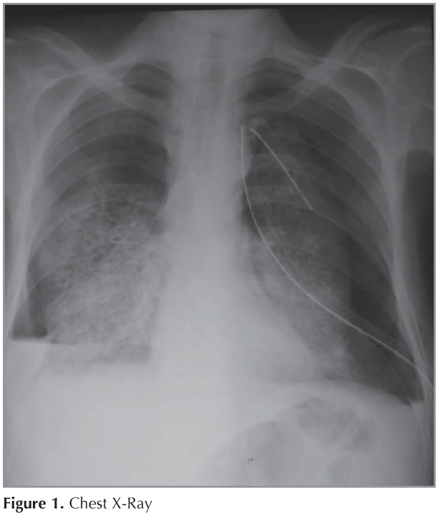
On the 20th day of anti-TB treatment, pneumothorax of left lung occurred. Chest tube thoracostomy was done. One day later, total pneumothorax of right lung occurred and right chest tube thoracostomy was done (Figure 1). Left and right sided pneumothorax significantly resorbed. But total pneumothorax of right lung recurred again. Right hemithorax was reexpanded later.Liver function tests and pancytopeni aim proved. The patient still remains drug free control.
Although miliary pattern and pneumothorax are rare radiological features in pulmonary TB, their incidences are nearly 1.3% and 1.5%, respectively (2). The diagnosis of pneumothorax may be delayed (3). The possible mechanism of pneumothorax in miliary TB is formation of small areas of confluent subpleural miliary nodules.
These nodules undergo caseous necrosis with subsequent rupture in to the pleural space. This will lead to air leak in to pleural cavity, causing pneumothorax (1).
Frequently pneumothorax is not seen at the beginning of therapy but is seen during the course of treatment when it is least expected (4,5). In the present case, bilateral pneumothorax occurred during anti-TB treatment. Right sided pneumothorax also recurred again. As a conclusion, although pneumothorax in miliary TB is rare, it can be recurrent. During anti-TB treatment, worsening should make us think of a pneumothorax.
CONFLICT of INTEREST
None declared.
REFERENCES
- Tasar MA, Bostanci I, Aslan S, Y?lmaz R, Dallar Y. Recurrent pneumothorax in an infant with miliary tuberculosis. Tuberk Toraks 2005;53:394-6.
- Akto?u S, Yorgancioglu A, Cirak K, K?se T, Dereli SM. Clinical spectrum of pulmonary and pleural tuberculosis: a report of 5,480 cases. Eur Respir J 1996;9(10):2031-5.
- Sharma N, Kumar P. Military tuberculosis with bilateral pneumothorax: A rare complication. Indian J Chest Dis Allied Sci 2002;44:125-7.
- Wammanda RD, Ameh EA, Ali FU. Bilateral pneumothoraxcomplication of miliary tuberculosis in children: Case report and review of literature. Ann Trop Paediatr 2003;23:149-52.
- Mert A, Bilir M, Akman C, Ozaras R, Tabak F, Ozturk R, et al. Spontaneous pneumothorax: A rare complication of military tuberculosis. Ann Thorac Cardiovasc Surg 2001;7:45-8.
Yaz??ma Adresi (Address for Correspondence)
Dr. Abdullah ??M?EK
Ankara Atat?rk G???s Hastal?klar? ve
G???s Cerrahisi E?itim ve Ara?t?rma Hastanesi,
G???s Hastal?klar? Klini?i,
ANKARA - TURKEY
e-mail: abdullahsimsek1@yahoo.com.tr