Hemotoraksla ba?vuran plevral multikistik mezotelyal proliferasyon olgusu
Ekrem ?ENT?RK1, Salih ?OKPINAR1, Serdar ?EN1, ?brahim METEO?LU2
1 Adnan Menderes ?niversitesi T?p Fak?ltesi, G???s Cerrahisi Anabilim Dal?, Ayd?n,
2 Adnan Menderes ?niversitesi T?p Fak?ltesi, Patoloji Anabilim Dal?, Ayd?n.
?ZET
Hemotoraksla ba?vuran plevral multikistik mezotelyal proliferasyon olgusu
Plevral multikistik mezotelyal proliferasyon ?ok nadir bir serozal patolojidir. Bu yaz?da dispne ve g???s a?r?s? ?ikayetiyle acil servise ba?vuran ve hemotoraksla prezente plevral multikistik mezotelyal proliferasyon olgusunu payla?mak istedik. Literat?rde plevral multikistik mezotelyal proliferasyon tan?mlanan bir olgu bildirilmi?tir. Plevral multikistik mezotelyal proliferasyon nadir bir benign patoloji olsa da hemotoraks gibi ya?amsal komplikasyonlara neden olabilir.
Anahtar Kelimeler: Plevral neoplazm, mezotelyal proliferasyon, hemotoraks.
SUMMARY
Pleural multicystic mesothelial proliferation that presented with hemothorax
Ekrem ?ENT?RK1, Salih ?OKPINAR1, Serdar ?EN1, ?brahim METEO?LU2
1 Department of Chest Surgery, Faculty of Medicine, Adnan Menderes University, Aydin, Turkey,
2 Department of Pathology, Faculty of Medicine, Adnan Menderes University, Aydin, Turkey.
Pleural multicystic mesothelial proliferation is a very rare serosal pathology. In this paper, we share a pleural multicystic mesothelial proliferation case arrives the emergency service with sudden chest pain and dyspnea complaint that presented with hemothorax complication. In the literature, there is only one pleural multicystic mesothelial proliferation issue that is determined by coincidence. Even though being a rare benign pathology; pleural multicystic mesothelial proliferation can cause some vital complications as a hemothorax.
Key Words: Pleural neoplasms, mesothelial proliferation, hemothorax.
Tuberk Toraks 2013; 61(1): 47-49 • doi: 10.5578/tt.2396
Geli? Tarihi/Received: 13/11/2012 - Kabul Edili? Tarihi/Accepted: 20/01/2013
INTRODUCTION
Multicystic mesothelial proliferation is a rare disease of the serosal cavity which is usually seen in abdominal region (1). Mesothelial cystic are classified in two groups which are pleuropericardial and pleural (2). Pleural multicystic mesothelial proliferation (PMMP) seen very rarely. The first case of pleural multicystic mesothelial proliferation was described by Ball in 1990 (3). In the literature, we coincidence with two PMMP case that one of them is multilocular.
PMMP, which is accepted as a benign pathology, is determined by coincidence and usually developed asymptomatic presentation. In this paper, we share a PMMP case with hemothorax complication.
CASE REPORT
A 43-year-old female patient arrive the emergency service with severe chest, chest pain and dyspnea and there is no respiration sound on right hemithorax upper zone. We do not detect any characteristics on the case's history. The other physical examination case, blood and biochemical investigation of the patient are normal. Airfluid level of upperright zone is watched in chest X-ray. In thorax computed tomography, hydropneumothorax is reported which is compatible with right lungupper lobe posterior segment (Figure 1A). We estimated a cyst hidatic with perforation complication.
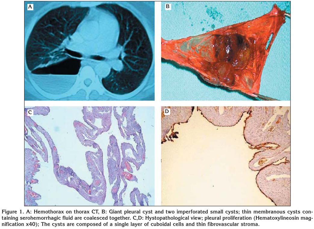
When the right hemithorax of thoracotomy with the diagnosis of perforated cyst was investigated, a perforated air cyst which has quite thin and transparent periphery was seen on right lung upper lobe posterior. In the air cyst, two imperforated cystic lesions with dark greenblackcolored liquid were monitored (Figure 1B). Also there was unretained dark-muddy hemorrhagic liquid (approximately; 2 liters) in the cyst and on thorax. The patient had a real hemothorax (blood in the pleural space) and bloodstained fluid inside the cyst (in the lung). Extra parenchymal cystic constitution is excised primarily. Histopathologic investigation is reported as compatible with PMMP (Figure 1C,D).
DISCUSSION
There is no consensus about the PMMP which is benign or malignant and the etiology of PMMP is not pointed. Therefore, definition of PMMP as histological lesions was found more suitable (1,4,5). Sometimes PMMP is being confused with malignant and due to the pleural liquid, it can be diagnosed, frequently, at the end of the clinical research (4). In our study, the patient arrive the emergency service with hemothorax complication. We think that the big cyst may be bleeding.
The rare PMMP has benign characteristics can be confused with malignant and can cause some emergent complications as in our case. In the literature, for the rare PMMP case, the only case is multilocular (5,6,7). There is limited number of pericardial cyst phenomenon series that is originated from mediastinum unilocular mesothelial in the literature (6,7,8). The pericardial or the other mediastinum mesothelial proliferation case is separated from PMMP via having unilocular structure and connective tissue and thought as mesothelial cyst (6,9).
The etiology of this disease is not known yet. However, in the etiology, unknown fibers and dusts generate inflammation mediators and mechanical injury in mesothelial cells and causes hyperplasic and neoplastic changes (6). According to some other scientist, tuberculosis pleurisy plays a part in the etiology. However, this relationship was not shown in the declared case. Some other scientists declare that tuberculosis pleurisy occurs as the result of migration of coelomic structure (10).
As a result, even though being a rare benign pathology; PMMP, can cause some vital complications as in our patient.
CONFLICT of INTEREST
None declared.
REFERENCES
- De Rosa G, Donofrio V, Boscaino A, Zeppa P, Staibano S. Multicystic mesothelial proliferation immunohistochemical, ultrastructural and DNA analysis of five cases. Virchows Arch A Pathol Anat 1992; 421: 379-85.
- Shields TW. Mesothelial and other less common cysts of the mediastinum. In: Shields TW (ed). General thoracic surgery. Philadelphia: Lippincott, Williams and Wilkins, 2000: 2423-7.
- Ball NJ, Urbanski SJ, Greet FHY, Kieser T. Pleural multicystic mesothelial proliferation: the socalled multicystic mesothelioma. Am J Surg Pathol 1990; 14: 375-8.
- Katoh S, Satoh M, Oouchi H, Imai S, Kusajima K. Pleural multicystic Mesothelial proliferation. Chest 1994; 105: 295-6.
- Pelosi G, Zannoni M, Caprioli F, Faccincani L, Battistoni MG, Balercia G, et al. Benign multicystic mesothelial proliferation of the peritoneum: immunohistochemical and electron microscopical study of a case and review of the literature. Histol Histopathol 1991; 6: 575-83.
- Psathakis K, Sampaziotis D, Psara A, Panagou P, Karameris A, and Tsintiris K. Pleural multicystic mesothelial proliferation. J Bronchol 2005; 12: 168-70.
- Cangemi V, Volpino P, Gualdi G, Polettini E, Frati R, Cangemi B, Piat G. Pericardial cysts of the mediastinum. J Cardiovasc Surg 1999; 40: 909-13.
- Salyer DC, Salyer WR, Eggleston JC. Benign developmental cysts of the mediastinum. Arch Pathol Lab Med 1977; 101: 136-9.
- Sasaki H, Yano M, Kiriyama M, Kaji M, Fukai I, Yamakawa Y, et al. Multicystic mesothelial cyst of the mediastinum: report of a case. Surg Today 2003; 33: 199-201.
- Lillie WI, McDonald JR, Clagett OT. Pericardial coelomic cysts and pericardial diverticula. J Thorac Surgery 1950; 20: 494.
Yaz??ma Adresi (Address for Correspondence):
Dr. Ekrem ?ENT?RK,
Adnan Menderes ?niversitesi T?p Fak?ltesi,
G???s Cerrahisi Anabilim Dal?,
09100 AYDIN - TURKEY
e-mail: ekremsenturk@hotmail.com