Endobron?iyal b?y?me g?steren akci?er adenokarsinomu
Katsunori Kagohashi, Koichi Kurishima, Hiroichi Ishikawa, Hiroaki Satoh
Tsukuba ?niversitesi Mito T?p Merkezi, ?? Hastal?klar? B?l?m?, Mito, Ibaraki, Japonya
?ZET
Endobron?iyal b?y?me g?steren akci?er adenokarsinomu
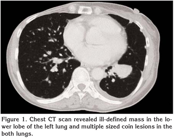
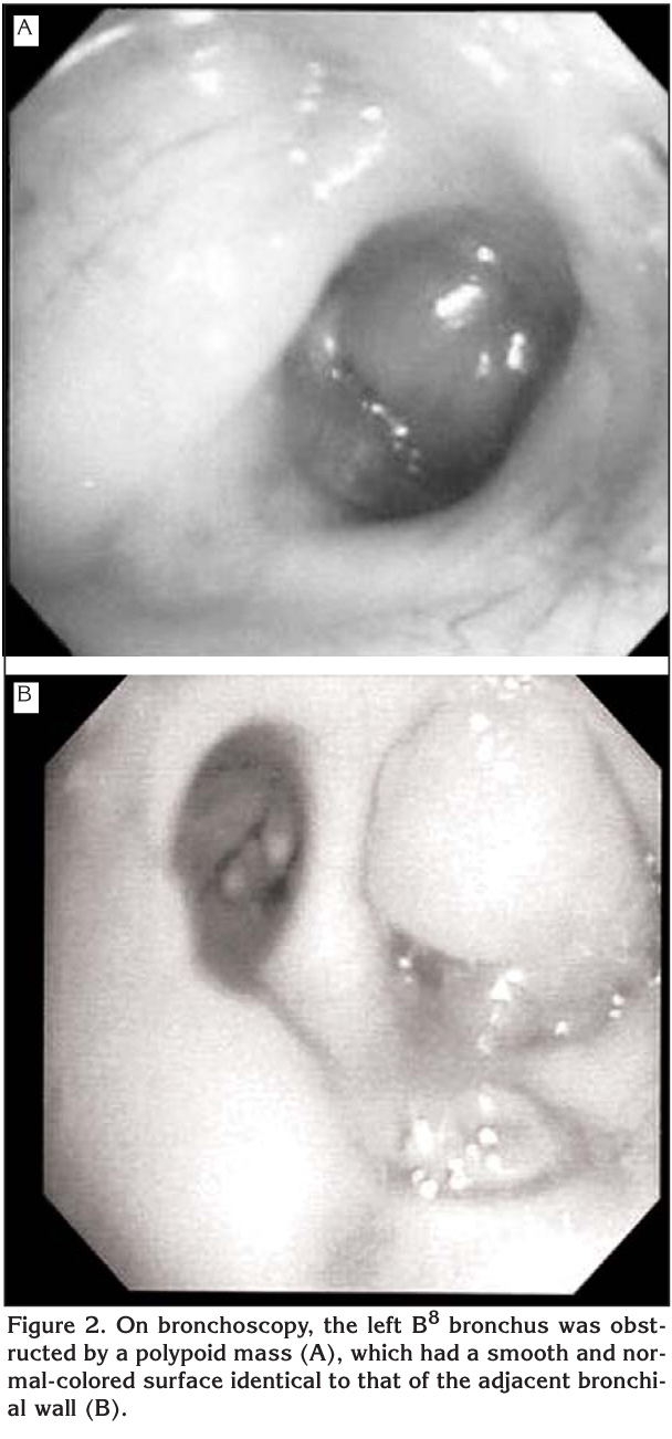
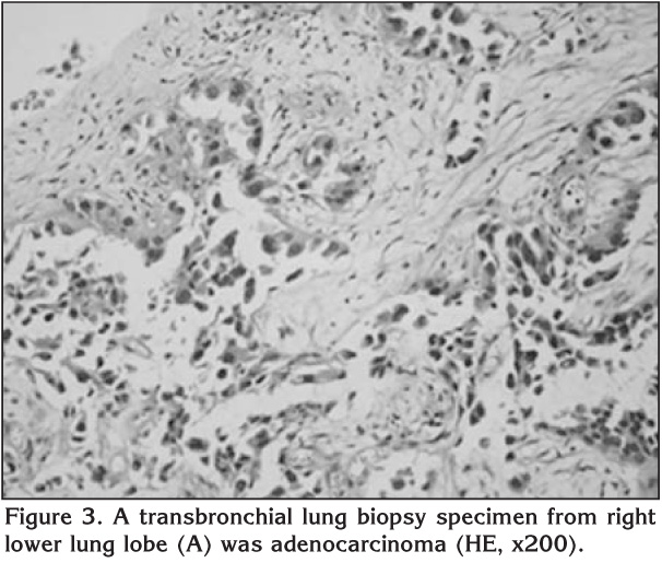
65 ya??nda, endobron?iyal b?y?me g?steren akci?er adenokarsinomlu nadir bir kad?n olguyu sunuyoruz. Toraks bilgisayarl? tomografide sol akci?er alt lobda kitle ve her iki akci?erde farkl? boyutlarda multipl nod?ller saptand?. Bronkoskopik incelemede sol B8 bron?ta endobron?iyal polipoid t?m?r izlendi. T?m?rden al?nan biyopsi sonucu akci?er adenokarsinom tan?s? konuldu. ?ok nadir olmas?na ra?men, bron?a kom?u pulmoner t?m?r ve endobron?iyal polipoid lezyonlu hastalarda akci?er adenokarsinomu ak?lda tutulmal?d?r.
Anahtar Kelimeler: Akci?er kanseri, adenokarsinom, endobron?iyal b?y?me.
SUMMARY
Lung adenocarcinoma with endobronchial growth
Katsunori Kagohashi, Koichi Kurishima, Hiroichi Ishikawa, Hiroaki Satoh
Department of Internal Medicine, Mito Medical Center, Tsukuba University, Mito, Ibaraki, Japan.
We report a rare case of lung adenocarcinoma with endobronchial growth in a 65-year-old woman. Chest computed tomography revealed an ill-defined mass in the lower lobe of the left lung and multiple sized nodular shadows in the both lungs.? An endobronchial polypoid tumor in the left B8 bronchus was found by bronchoscopic examination.? A biopsy specimen obtained from the tumor diagnosed lung adenocarcinoma.? Although very rare, we should therefore keep in mind that patients who have a pulmonary tumor adjacent to the bronchus with an endobronchial polypoid lesion may have lung adenocarcinoma.
Key Words: Lung cancer, adenocarcinoma, endobronchial growth.
Although adenocarcinoma is one of the most common histologic types of primary lung cancer, endobronchial extension is very rare (1,2). Clinical differential diagnosis of endobronchial lesion may include a variety of conditions including non-malignant tumors, primary lung carcinoma other than adenocarcinoma, and endobronchial metastasis of carcinoma from extrapulmonary organs (3,4,5,6,7,8). We report herein a patient of lung adenocarcinoma with endobronchial growth.
Case Report
A 65-year-old female was referred to our hospital due to multiple sized nodular shadows in both lungs. Nineteen years previously she had mastectomy for breast cancer and 5 years previously she had thyroidectomy for thyroid cancer. On admission, laboratory examination revealed a hemoglobin of 12.5 g/dL, hematocrit of 37.2%, and a C-reactive protein count of 0.64 mg/dL. Serum level of carcinoembryonic antigen was elevated to 156.7 ng/mL. X-ray and computed tomography (CT) of the chest revealed ill-defined mass in the lower lobe of the left lung and multiple and various sized coin lesions in the both lungs (Figure 1). On bronchoscopy, the left B8 bronchus was obstructed by a polypoid mass, which had a smooth and normal-colored surface identical to that of the adjacent bronchial wall (Figure 2). Transbronchial biopsy revealed the tumor to be adenocarcinoma (Figure 3). Immunohistochemical examination revealed positive for cytokeratin (CK)-7, CK-20, thyroid transcription factor 1 (TTF-1), and surfactant protein A (SP-A). Brain magnetic resonance imaging, abdominal ultrasonography, bone scintigraphy did not reveal malignancy. The tumor was diagnosed as primary lung carcinoma with a clinical stage of T2N2M1-stage IV. She was treated with two courses of amrubicin and has been in good condition for 8 months since the initiation of the chemotherapy.
Discussion
The radiolographic findings due to endobronchial lesion are considerably variable. Lobar or segmental atelectasis and pneumonic infiltration are commonly observed. The radiological differential diagnosis of the endobronchial mass lesion includes non-malignant tumors, primary lung carcinoma, and endobronchial metastasis of carcinoma from extrapulmonary organs (3,4,5,6,7,8). In addition, the mass-like pulmonary opacity, which probably represents endobronchial infectious process such as mucus plugs distal to a centrally obstructing lesion due to actinomycosis, aspergillosis, or tuberculosis also simulate a endobronchial mass on CT scan (9,10,11). Squamous cell carcinoma is the most common histologic type of primary lung carcinoma in central location and endobronchial extension (4). In this cell type, polypoid lesion with a rough surface covered with necrotic material is the most predominant finding (12,13). Breast, kidney, and colon are three of the most common sites which produce endobronchial metastasis (5,6,7). Among them, metastatic endobronchial tumor originating from colorectal carcinoma often presents as a polypoid or nodular lesion covered with necrotic material (14). On the other hand, non-malignant endobronchial tumors usually have smooth surface with uniform color (15). It is generally accepted that bronchoscopic findings of endobronchial lesion of metastasis is not easily distinguishable from those of primary lung carcinoma and non-malignant tumor (16,17). Diagnosis of endobronchial lesions can be made by bronchoscopic examination because most lesions are within the view and grasp of the bronchoscopic field. Therefore, bronchoscopic biopsy is the key to making an accurate and definitive diagnosis. In some cases, the value of bronchoscopic examination may be limited because of the admixture of necrotic material interfering the opportunity to obtain a proper diagnostic specimen (9,10,11), however, the pathological diagnosis using specimens obtained by bronchoscopic biopsy is mandatory for a correct diagnosis.
Adenocarcinoma is a most common histologic type of primary lung carcinoma and is mostly peripheral location. Even in cases of bronchial involvement, submucosal extension and narrowing of the bronchial lumen are usually seen (18). In small parts of patients, therefore, the presenting symptoms are cough, hemoptysis, and less frequently, pulmonary infections. In large parts of them, the patients even in advanced disease have no pulmonary symptoms, and the lesion was discovered incidentally on routine radiological examination. Although very rare, there were some cases with lung adenocarcinoma with endobronchial growth (1,2). Murata et al. reported a case of lung poorly differentiated adenocarcinoma with endobronchial growth from the periphery towards the hilum (1). Kodama et al. described 5 cases of lung adenocarcinoma with predominantly endobronchial polypoid growth (2). Our patient had a history of completely resected breast and thyroid carcinomas and she had no recurrence. Both breast and lung carcinomas are adenocarcinoma, but we diagnosed this endobronchial tumor to be a primary lung adenocarcinoma because immunohistochemical examination revealed positive for CK-7, CK-20, TTF-1 and SP-A. In our patient, as observed in a patient reported by Kodama et al., primary adenocarcinoma originated from left lower lobe of the lung endobronchially extended towards the left lower bronchus (2). On bronchoscopic examination, it was an irregularly surface polypoid lesion with no necrotic material coverage. Bronchoscopically, differential diagnosis from metastatic endobronchial tumor could not be made because the lesion had no specific finding.
Although it is very rare tumor presentation, we should always be considered that lung adenocarcinoma can develop endobronchial growth. In addition, this report confirms the importance of bronchoscopical examination in patients with a history of extrapulmonary malignancy who have endobronchial manifestations.
CONFLICT of INTEREST
None declared.
REFERENCES
- Murata Y, Inomata T, Yamamoto A, Yamashiro T, Moriki T, Yoshida S. Endobronchial growth patterns in peripheral adenocarcinoma of the lung. J Thorac Imaging 2002; 17: 89-91. [?zet]
- Kodama T, Shimosato Y, Koide T, Watanabe S, Yoneyama T. Endobronchial polypoid adenocarcinoma of the lung. Histological and ultrastructural studies of five cases. Am J Surg Pathol 1984; 8: 845-54. [?zet]
- Unger M. Endobronchial therapy of neoplasms. Chest Surg Clin North Am 2003; 13: 129-47. [?zet]
- Koss MN, Hochholzer L, Frommelt RA. Carcinosarcomas of the lung: a clinicopathologic study of 66 patients. Am J Surg Pathol 1999; 23: 1514-26. [?zet]
- Katsimbri PP, Bamias AT, Froudarakis ME, Peponis IA, Constantopoulos SH, Pavlidis NA. Endobronchial metastases secondary to solid tumors: report of eight cases and review of the literature. Lung Cancer 2000; 28: 163-70. [?zet]
- Braman SS, Whitcomb ME. Endobronchial metastasis. Arch Intern Med 1975; 135: 543-7. [?zet]
- Seo JB, Im JG, Goo JM, Chung MJ, Kim MY. Atypical pulmonary metastases: spectrum of radiologic findings. Radiographics 2001; 21: 403-17. [?zet] [Tam Metin] [PDF]
- Shah H, Garbe L, Nussbaum E, Dumon JF, Chiodera PL, Cavaliere S. Benign tumors of the tracheobronchial tree. Endoscopic characteristics and role of laser resection. Chest 1995; 107: 1744-51. [?zet] [PDF]
-
Lau KY. Endobronchial actinomycosis mimicking pulmonary neoplasm. Thorax 1992; 47: 664-5.
[?zet] [PDF] - Kim JS, Rhee Y, Kang SM, Ko WK, Kim YS, Lee JG, et al. A case of endobronchial aspergilloma. Yonsei Med J 2000; 41: 422-5. [?zet]
- Van den Brande P, Lambrechts M, Tack J, Demedts M. Endobronchial tuberculosis mimicking lung cancer in elderly patients. Respir Med 1991; 85: 107-9. [?zet]
- Murakami S, Watanabe Y, Saitoh H, Yamashita R, Shimizu J, Oda M, et al. Treatment of multiple primary squamous cell carcinomas of the lung. Ann Thorac Surg 1995; 60: 964-9. [?zet] [PDF]
- Saida Y, Kujiraoka Y, Akaogi E, Ogata T, Kurosaki Y, Itai Y. Early squamous cell carcinoma of the lung: CT and pathologic correlation. Radiology 1996; 201: 61-5. [?zet]
- Oshikawa K, Ohno S, Ishii Y, Kitamura S. Evaluation of bronchoscopic findings in patients with metastatic pulmonary tumor. Intern Med 1998; 37: 349-53. [?zet]
- Wilson RW, Kirejczyk W. Pathological and radiological correlation of endobronchial neoplasms: Part I, Benign tumors. Ann Diagn Pathol 1997; 1: 31-46. [?zet]
- Lam B, Wong MP, Fung SL, Lam DC, Wong PC, Mok TY, et al. The clinical value of autofluorescence bronchoscopy for the diagnosis of lung cancer. Eur Respir J 2006; 28: 915-9. [?zet] [Tam Metin] [PDF]
- Shulman L, Ost D. Advances in bronchoscopic diagnosis of lung cancer. Curr Opin Pulm Med 2007; 13: 271-7. [?zet]
- Schenk DA, Bryan CL, Bower JH, Myers DL. Transbronchial needle aspiration in the diagnosis of bronchogenic carcinoma. Chest 1987; 92: 83-5. [?zet] [PDF]
Yaz??ma Adresi (Address for Correspondence):
Dr. Hiroaki Satoh,
Department of Internal Medicine,
Mito Medical Center, Tsukuba University, Mito, Ibaraki,
310-0015, IBARAKI - JAPAN
e-mail: hirosato@md.tsukuba.ac.jp